
The cardiac activity is normally monitored by an implantable stimulation device (pacemaker) through the intracavitary detection of the electric signals associated with atrial and ventricular depolarization (A and R waves, respectively) .
The electric signals, however, provide information limited to atrial and/or ventricular timing. For an optimal control of the heart – pacemaker system, the electrical sensing should better be integrated with the assessment of mechanical cardiac activity and related hemodynamic parameters.
The mechanical activity can be assessed by using specific sensors, or alternatively through electrical impedance measurement, which can be performed by means of the same catheters commonly applied in cardiac stimulation, without any reduction in performance and precision with the progress of time. Indeed, the structural and geometric modifications taking place during the cardiac cycle result in periodic impedance fluctuation, which can represent an index of ventricular contractility and systolic outflow.
A new option in cardiac impedance measurement is the Trans-Valvular Impedance (TVI) technique.
TVI is derived between right atrium and ventricle in standard dual-chamber implants (DDD or VDD with single-pass lead).
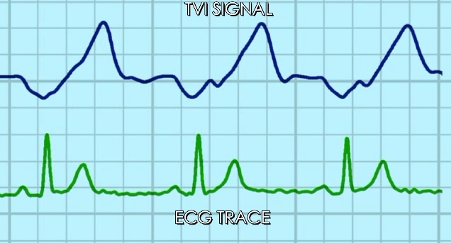
As shown in the tracings, TVI decreases during the atrial systole and the diastolic passive filling, and increases during the ventricular systole, reaching the maximum peak at the end of the Q-T period (i.e.: at the end of ventricular ejection).
The increasing clinical experience, including thousands of implants performed in the main International Centers, confirms the correlation between TVI, cardiac mechanics, and corresponding hemodynamic performance.
This allows innovative important applications, like the ejection surveillance at every paced or sensed cardiac beat, and the regulation of stimulation timing. Furthermore, the Cardiologist can take advantage of TVI indications in the preliminary evaluation of the hemodynamic stability of pacemaker patients.
One step beyond in the hemodynamic regulation of cardiac pacing:
TVI signal automatic calibration

Autosetting of TVI recording configuration, with immediate availability of TVI-dependent functions.
Ejection check after ventricular pacing
The TVI signal is applied to confirm the actual occurrence of ejection after a ventricular pacing pulse. In the event of missed ejection, the pulse energy is increased to restore effective stimulation.

If the expected TVI increase is not recorded after a pacing pulse, failure of ventricular capture is assumed and the pulse energy is increased.
The pacemaker stores in memory the number of no capture alarms and the trend of pulse parameters.
Ejection Check After Ventricular Sensing
The TVI signal is applied to confirm the actual occurrence of ejection after a ventricular sensing event. In case of missed ejection, the pacing mode is switched over from any ventricular-inhibited to the corresponding ventricular-triggered mode, in order to prevent the risk of false inhibition induced by oversensing.

If the expected TVI increase is not recorded after a series of 3 ventricular sensing events, any V-inhibited pacing mode is switched over to the corresponding V-triggered one, in order to ensure safety stimulation.
V-triggered and inhibited pacing are alternated at every beat, so that the programmed pacing mode is re-established as soon as a reliable sensing function is restored.
The number of no-ejection alarms after ventricular sensing is stored in memory.
Promotion of intrinsic AV conduction warranted by TVI recording

TVI-based ejection surveillance enables dual chamber pacing after a single event of false ventricular inhibition.
Long-term TVI signal comparison
Acquired waveform comparison with the one at previous check and the one selected as starting reference.

- TVI waveform comparison to assess long-term hemodynamic stability
- Automatic measurement of basic parameters
- Diastolic and systolic TVI trends
…allow screening patients at risk for heart failure
Full papers in details:
![]() Acute effects of right ventricular pacing on cardiac haemodynamics and transvalvular impedance.
Acute effects of right ventricular pacing on cardiac haemodynamics and transvalvular impedance.
Taborsky M, Fedorco M, Skala T, Kocianova E, Pastucha D, Richter D, Petrkova J, Di Gregorio F, Barbetta A, Vaclavik J
Biomedical Papers (2013) 157, in press.
![]() Pre-print Link: click here >>
Pre-print Link: click here >>
![]() Left ventricular mechanical activity detected by impedance recording
Left ventricular mechanical activity detected by impedance recording
Taborsky M, Kupec J, Vopalka R, Barbetta A, Di Gregorio F
Europace (2010) 12, 534–539
![]() Link Full paper: click here >>
Link Full paper: click here >>
![]() Rate-responsive pacing regulated by cardiac haemodynamics
Rate-responsive pacing regulated by cardiac haemodynamics
Gasparini G, Curnis A, Gulizia M, Occhetta E, Corrado A, Bontempi L, Mascioli G, Francese GM, Bortnik M, Magnani A, Di Gregorio F, Barbetta A, Raviele A
Europace (2005) 7, 234–241
![]() Link Full paper: click here >>
Link Full paper: click here >>


Products | Technology | Support | Contacts